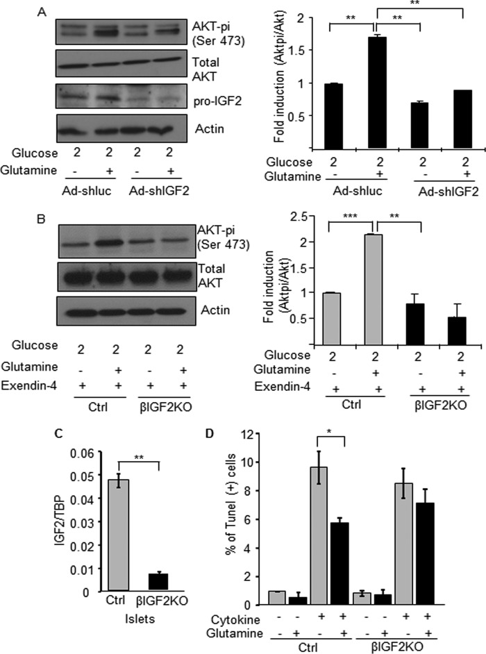
FIGURE 10.
Glutamine-induced IGF2 secretion leads to enhanced Akt signaling and protection against apoptosis. A, MIN6 cells were transduced with igf2-specific or unrelated (luc) shRNAs expressing adenoviruses. 48 h later, the cells were preincubated for 2 h in 2 mm glucose then for 3 h in 2 mm glucose with or without 2 mm glutamine. Western blot analysis of Pi-Akt, total Akt, pro-IGF2, and actin is shown on the left panel. Right panel: quantification of Pi-Akt/Akt ratio. B, islets from control (Ctrl) and beta cell-specific Igf2KO mice (βIgf2KO) were pretreated with exendin-4 for 18 h to increase IGF1R expression and then incubated as the MIN6 cells were described in A). Left: Western blot analysis of Pi-Akt, Akt, and actin. Right: quantification of the results. C, quantitative PCR analysis of Igf2 mRNA expression in islets from control and βIgf2KO mice. D, apoptosis measured in islet cells from control (Ctrl) and beta cell-specific Igf2KO mice (βIgf2KO) exposed, as indicated, to cytokines and glutamine for 16 h before TUNEL analysis. The data are the means ± S.D. from three independent experiments. *, p < 0.05; **, p < 0.01; ***, p < 0.001.

