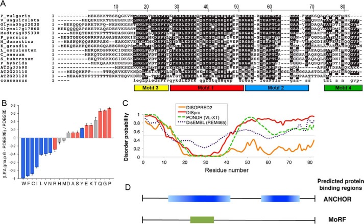
FIGURE 1.
In silico analysis of the PvLEA6 protein sequence. A, alignment of group 6 LEA proteins showing the conserved regions and distinctive motifs of the family. B, comparative analysis of amino acid composition between group 6 LEA proteins and a subset of ordered proteins amenable to crystallization studies (PDBS25) using Composition Profiler software (31). Red, blue and gray bars refer to amino acid residues promoting disorder, order, or neutrality, respectively. C, prediction of ordered/disordered regions in PvLEA6 protein using different disorder predictors as indicated. Numbers in the x axis correspond to those in the upper line in A. D, putative protein binding regions in PvLEA6 protein predicted using ANCHOR (32) and MoRFpred (33).

