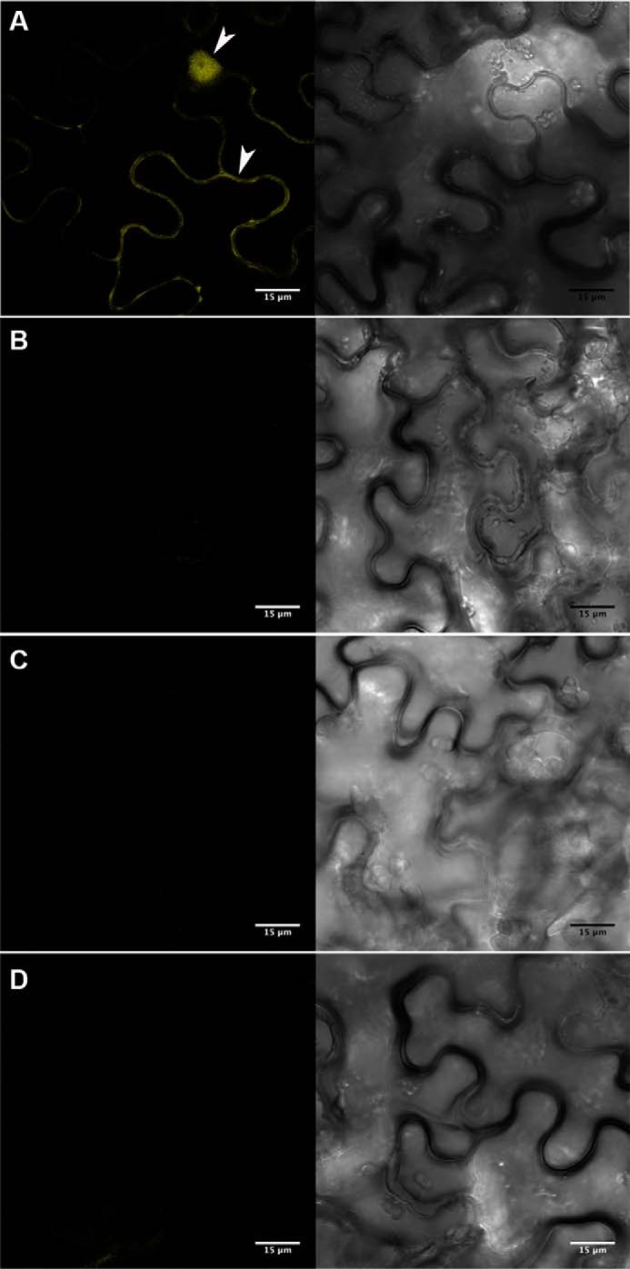
FIGURE 11.

Visualization of PvLEA6 dimer using BiFC assay in N. benthamiana epidermis cells. Complete PvLEA6 ORF was cloned in BiFC vectors (27). Transient expression was analyzed by confocal microscopy. A, PvLEA6 dimers were detected in cytoplasm and nuclei of epidermal cells co-transformed with YFPN43-PvLEA6 and YFPC43-PvLEA6. White arrowheads indicate cytosol and nuclear signals. Cytosol is pressed by a large central vacuole against cell membrane. No fluorescence was detected in controls. B, pYFPN43 and pYFPC43, empty vectors. C, YFPN43-PvLEA6 and pYFPC43. D, pYFPN43 and YFPC43-PvLEA6. Scale bar corresponds to 15 μm. Both images within A–D correspond to the same field. Images obtained with UV light are shown at the left side and with white light at the right side.
