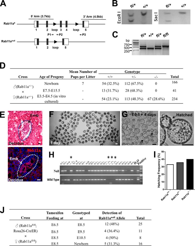
FIGURE 1.
Rab11a global knockout mice die at the peri-implantation stage. A, schematics of Rab11aflox and Rab11anull alleles. P1, P2, and P3 represent genomic PCR primers to identify specific alleles. B, Southern blots confirmed correctly targeted ES cell clones. C, PCR using the primer set P1 and P2 confirmed the derivation of Rab11afl/fl mice. D, summary of genotyping results of 166 neonatal pups and 275 embryos produced from intercrossing Rab11a+/− mice. 67 live Rab11anull embryos were identified from in vitro cultured blastocysts. No live null embryos or pups were detected in utero or after birth. E, representative implanted embryos (Emb), all of which were Rab11a-positive wild types. H&E and Rab11a staining of uterus sections is shown. Scale bars = 10 μm. F, fertilized eggs (E0.5) from Rab11a+/− mating were cultured for 1 day in vitro. Scale bar = 50 μm. G, after 4 days of development in culture, Rab11anull blastocysts underwent hatching (arrows). Scale bars = 40 μm. H, genotyping of individual hatched blastocysts identified Rab11anull embryos (asterisks). I, Rab11anull embryos demonstrated equivalent hatching activity as wild types. J, Rab11a was inducibly deleted from E6.5 or E8.5 embryos via tamoxifen-gavaging pregnant Rab11afl/fl females, which were plugged by Rab11afl/fl;Rosa26-CreER males. A summary of the genotyping results showed the positive detection of null alleles from live embryos collected at the designated time points.

