FIGURE 3.
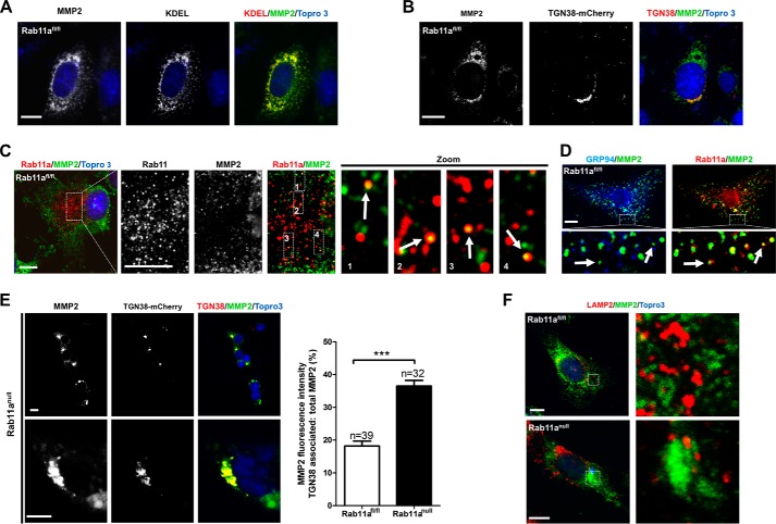
Rab11a vesicles intersect MMP2 vesicular export. A and B, confocal immunofluorescent staining of wild-type MEFs for endogenous MMP2 (green) showed that the large majority of MMP2 was in the ER (red, A, KDEL), whereas a small portion of MMP2 (green) was localized in the TGN (red, B, TGN38-mCherry). Scale bars = 10 μm. KDEL-EGFP was pseudocolored as red. KDEL, ER retention motif with amino acid sequence as lysine-aspartic acid-glutamic acid-leucine. C, confocal immunofluorescent staining of wild-type MEFs for endogenous MMP2 (green) and Rab11a (red) showed that a small fraction of MMP2+ vesicles were positive for Rab11a. Scale bars = 10 μm. D, confocal immunofluorescent staining of wild-type MEFs for endogenous GRP94 (blue), MMP2 (green), and Rab11a (red) showed Rab11a-positive, ER-negative MMP2 vesicles. Scale bar = 10 μm. E, confocal immunofluorescent staining of Rab11anull MEFs for endogenous MMP2 (green) and TGN (red, TGN38-mCherry) showed a clear aggregation of MMP2 in the TGN. Note that all Rab11anull MEF cells displayed an identical MMP2 aggregation pattern regardless of whether or not they received a TGN38-mCherry transfection. Untransfected cells are indicated by arrows. The bar graph shows the percentages of TGN-associated MMP2 in wild-type versus Rab11anull MEFs. ***, p < 0.001. Scale bars = 10 μm. F, confocal immunofluorescent staining of wild-type and Rab11anull MEFs for endogenous MMP2 (green) and LAMP2 (red). No colocalization was detected in either cell type. Scale bars = 10 μm.

