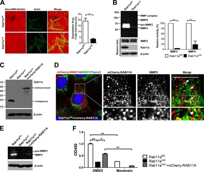
FIGURE 4.
Rab11a ablation impairs MMP2 secretion in MEFs. A, extracellular matrix degradation assays showed reduced gelatin degradation by Rab11anull MEFs (bottom row), compared with wild-type MEFs (top row). Degradation areas were quantified from three independent experiments. ***, p < 0.001. B, in-gel zymography assays showed reduced MMP2 and MMP9 secretion by Rab11anull MEFs. Shown are quantified zymography measurements of MMP2 and MMP9 from three independent experiments. ***, p < 0.001. C, Western blot analyses detected the expression of exogenous mCherry-RAB11A in stably infected Rab11anull MEFs. D, confocal immunofluorescent staining of mCherry-RAB11A rescued Rab11anull MEFs. Colocalization of some endogenous MMP2 vesicles (green) with mCherry-RAB11A (red) was detected (arrows). Scale bars = 10 μm. E, zymography assays using the mCherry-RAB11A rescued Rab11anull MEFs showed partially restored MMP2 activities. F, MMP2-specific ELISA showed a decreased MMP2 secretion by Rab11anull MEFs compared with wild-type MEFs. Monensin also inhibited MMP2 secretion in wild-type MEFs. mCherry-RAB11A-rescued Rab11anull MEFs showed a partial restoration of MMP2 secretion, which remained sensitive to monensin blockage. Data represent two independent experiments. *, p < 0.05; **, p < 0.01. DMSO, dimethyl sulfoxide.

