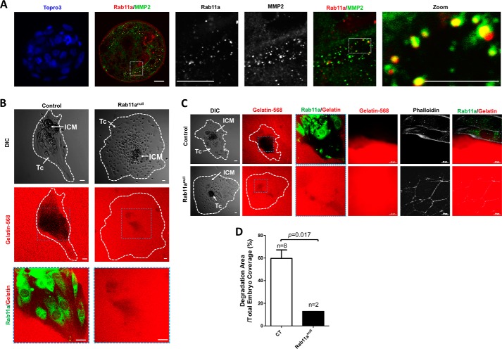
FIGURE 6.
Impaired gelatin degradation by Rab11anull blastocysts. A, confocal immunofluorescent staining on wild-type blastocysts showed colocalization between MMP2+ vesicles (green) and Rab11a (red). Scale bars = 10 μm. B and C, individual blastocysts (hatched) were seeded on chamber slides precoated with gelatin 568 (red). Extracellular matrix degradation activities, indicated by the loss of red fluorescent signals, by individual blastocysts were measured 3 days after embryo seeding. Rab11a staining (green) was used to identify null embryos. The leading edges of the embryo C were outlined by phalloidin 350 staining. Scale bars = 20 μm. DIC, differential interference contrast. D, the percentage of degradation areas were quantified and compared between wild-type and Rab11anull blastocysts. Tc, trophoblastic cells; ICM, inner cell mass.

