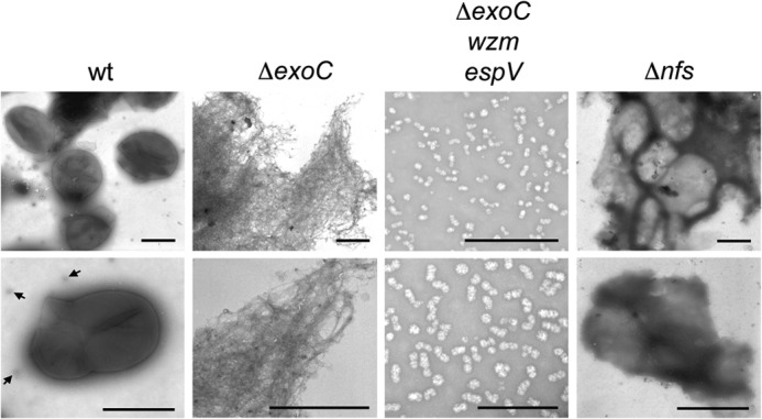
FIGURE 1.

Negative stain electron microscopy of material isolated during spore coat purification. Wild type (wt; strain DK1622), ΔexoC (PH1261), ΔexoC wzm epsV (PH1296), and ΔnfsA-H (Δnfs; PH1200) cells were induced to sporulate for 4 h by addition of glycerol and subjected to a spore coat isolation protocol. Representative pictures at lower (top panels) and higher (bottom panels) magnifications are shown. Bar corresponds to 1 μm for WT, ΔexoC, and ΔnfsA-H panels, and 500 nm for ΔexoC wzm epsV panels. Arrows point to particles in the wild type similar to those isolated from the ΔexoC wzm epsV mutant.
