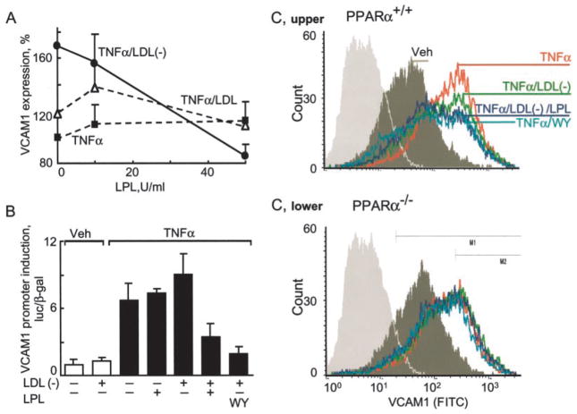
Fig. 1. LPL treatment of LDL(−) opposes VCAM-1 induction in a PPARα-dependent manner.
A, confluent ECs were pretreated with the amount of LPL indicated and total LDL or LDL(−) (10 μg/ml, 3 h) and then stimulated with human TNFα (10 ng/ml, 12 h). VCAM1 expression, measured by ELISA, is shown as a percentage of VCAM1 induced by TNFα alone. Mean ± S.D. of triplicate experiments is shown. B, BAECs were transfected with a human VCAM1 promoter construct as described under “Experimental Procedures.” Cells were pretreated (4 h) with the synthetic PPARα ligand WY14647 (WY; 250 μM) or LPL (200 units/ml) in the presence and absence of LDL(−) (10 μg/ml) and then stimulated with human TNFα (10 ng/ml, 12 h). VCAM1 promoter activation is shown as a comparison between stimulated and non-stimulated cells. In all transfection experiments, luciferase activity was normalized to co-transfected β-galactosidase activity. Data represent mean ± S.D. of triplicate measurement of experiments performed three times using pooled LDL(−) from multiple normal donors. Veh, vehicle. C, fluorescence-activated cell sorter analysis of VCAM1 expression on ECs isolated from the hearts of PPARα+/+ and PPARα−/− mice. Confluent ECs were pretreated with WY14647 (WY; 250 μM), the indicated amounts of LPL and LDL or LDL(−) (10 μg/ml 3 h), and stimulated with murine TNFα (50 ng/ml, 12 h). FITC, fluorescein isothiocyanate.

