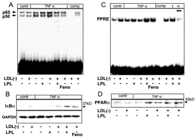
Fig. 2. LPL treated LDL(−) inhibits NFκB activation and increases IκBα expression while inducing PPRE binding.
Confluent human ECs were pretreated with LPL (200 units/ml, 4 h) in the presence or absence of LDL(−) (10 μg/ml) and then stimulated with human TNFα (10 ng/ml, 1.5 h). Fenofibric acid (Feno; 100 μM) pre-treatment was used for comparison. Nuclear extracts were analyzed by EMSA using 5 μg of nuclear extracts and 100 ng of radiolabeled NFκB (A), AP-1 (data not shown), and PPRE (C) sequences (Santa Cruz Biotechnology). Supershift analysis was performed to confirm the identity of PPARα and PPARγ as well as p65 and p50 (data not shown) using specific antibodies. Cytosolic fractions (25 μg protein) of the same cells were analyzed for IκBα expression (B), whereas nuclear fractions (25 μg protein) were studied for PPARα expression (D) using Western blotting. Glyceraldehyde-3-phosphate dehydrogenase (GAPDH) expression confirmed equal loading. Data shown are representative of one experiment from three with similar results.

