Figure 1.

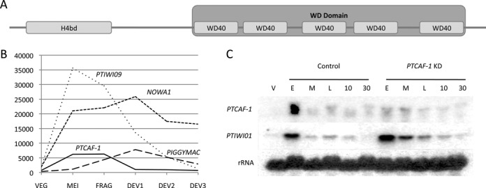
PtCAF-1 structure and expression pattern. (A) Schematic drawing of the PtCAF-1 domain architecture. The 391 amino acid long protein has both a histone binding domain (H4bd) between amino acids 15 and 84 and five WD40 repeats between the amino acids 152 and 370. The five WD40 repeats fold to form a WD domain. (B) Expression of PTCAF-1 based on microarray data from (29,36) during sexual reproduction of Paramecium tetraurelia in comparison to NOWA1, PTIWI09 and PIGGYMAC. VEG: vegetative cells; MEI: beginning of macronuclear fragmentation and micronuclear meiosis; FRG: population in which about 50% of cells have a fragmented old macronucleus; DEV1: earliest stage at which a significant proportion of cells has visible macronuclear anlagen; DEV2: majority of cells with macronuclear anlagen; DEV3: population of cells 10 h after DEV2. (C) Expression of PTCAF-1 and PTIWI01 analysed by northern blot. Cells during vegetative growth (V) and during autogamy where sampled from control and PTCAF-1 KD cultures and analysed for PTCAF-1 and Ptiwi01 expression. E indicates a culture with 50% of cells with fragmented MAC (E ≈ FRG in B). M indicates a culture with 100% of cells with fragmented MAC (M ≈ between FRG and DEV1 in B). L indicates a culture 6 h after M (L ≈ DEV1 in (B)). 10 and 30 indicate cultures 10 and 30 h after M, respectively (10 ≈ DEV2 in B, 30 is ∼10 h later than DEV3 in B). Consecutive hybridizations on one membrane were performed with PTCAF-1, PTIWI01 and 17S rRNA probes.
