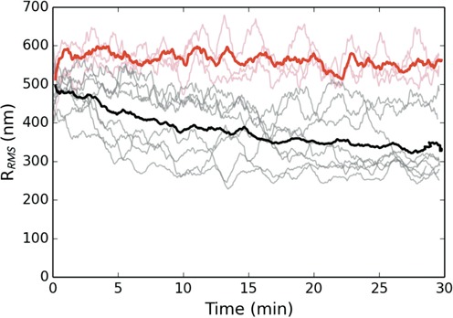
Figure 5.

Dynamics of DNA bound by 200 nM of H-NS and 2 mM of MgCl2 as a function of time. The black curves display the RMS excursion of the tethered microspheres as a function of time after release from the optical trap. The red curves are a control absent an applied force. The prominent solid curves are averaged over the respective trajectories for each population. All trajectories have been smoothed by a 60 s running window. Notice that the DNA, bound by H-NS alone, can be triggered to collapse in the presence of millimolar concentrations of Mg2+.
