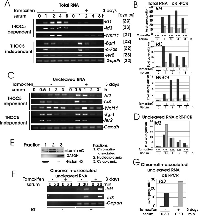
Figure 2.

Upon depletion of THOC5, Id1, Id3 and Wnt11 genes were transcribed, but not released from chromatin. (A and B) ERT2Cre THOC5 (flox/flox) MEF cells were treated with or without tamoxifen for 2 days prior to incubation in medium without serum for 24 h. The cells were then stimulated with serum for 0, 1, 2, 4 and 8 h. Total RNA was isolated from each samples and semi-quantitative RT-PCR (A) and quantitative (q)RT-PCR (B) were performed. Primers were located in different exons (Table 1). (C and D) Cells were treated as described in (A) but stimulated with serum for 0.5, 1, 2 and 3 h. Nuclear RNA were isolated and used for RT-PCR. Primers were shown in Table 1. (E) Proteins were extracted from chromatin associated nucleoplasmic and cytoplasmic fractions and were used for LaminAC, GAPDH and Histone H3-specific immunoblot. (F and G) Chromatin associated RNAs were isolated from cells stimulated for 30 min with serum in the presence (Tamoxifen−) or absence (Tamoxifen+) of THOC5 and RT-PCR was performed using the same primers as (C and D). Three independent experiments were performed.
