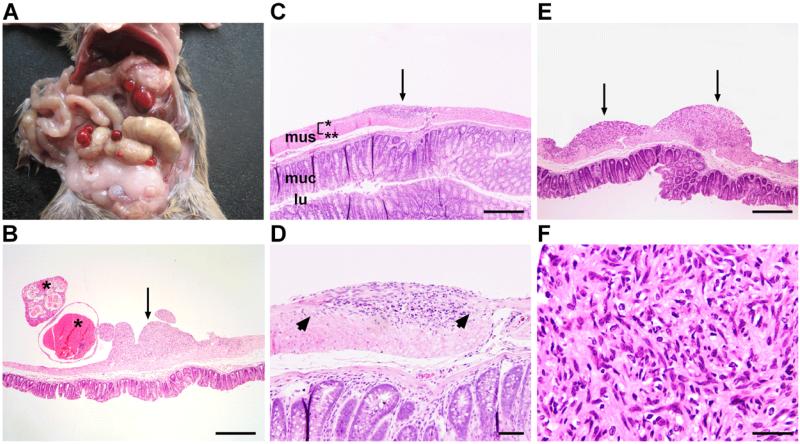
Figure 1. Ptch mutant mice develop gastrointestinal tumors.
(A) Gross appearance of tumors of Ptchflox/floxLysMcre+/− mice. (B) Tumors either have a solid (arrow) or cystic appearance (asterisks). The cystic appearance is associated with intratumoral bleeding. (C) Shows a precursor lesion (arrow). mus, muscularis (*: longitudinal muscle layer; **: circular muscle layer); muc, mucosa; lu, lumen of the intestine. (D) Arrows point to the intact myenteric plexus. (E) When precursor lesions become larger (arrows) they adopt a (F) GIST-like histology. Scale bars in μm: (B,C,E) 500; (D) 100; (F) 50.

