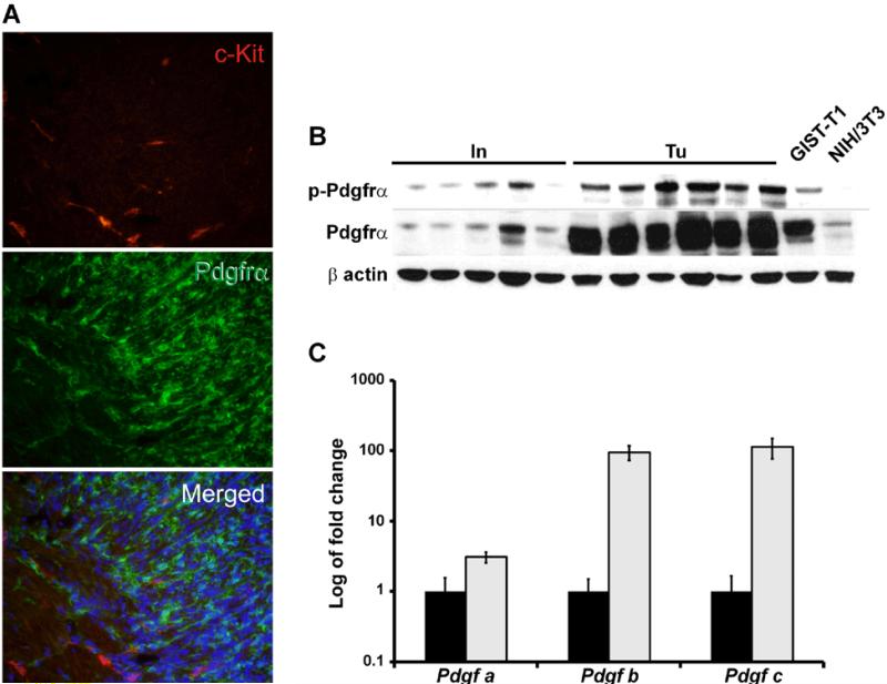
Figure 4. Tumors of Ptch mutant mice express Pdgfrα but not Kit.
(A) Immunofluorescence analysis of tumors of Ptchflox/floxLysMcre+/− mice using an anti-Pdgfrα (green) and anti-Kit antibody (red). No Pdgfrα/Kit double-positive cells were detected in the tumors. Scale bar 50 μm. (B) Western Blot analysis of tumors (Tu) and normal small intestine (In) of Ptchflox/floxLysMcre+/− mice using anti-Pdgfrα and anti-pPdgfrα (phosphorylated Pdgfrα) antibodies. GIST-T1 and NIH/3T3 cells were used as control samples. (C) qRT-PCR analysis of Pdgf-a, b and c in normal intestine and in tumors. Expression levels in tumors are shown in relation to normal intestine, which was set=1. (black bars: intestine; grey bars: tumors).

