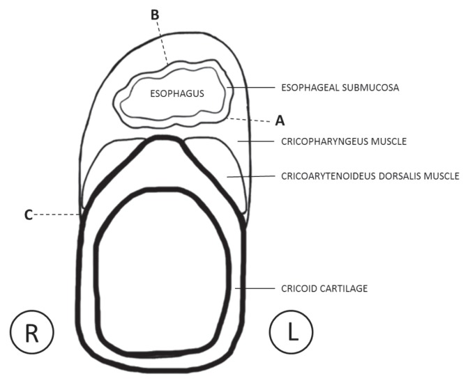
Figure 2.
Line drawing depicting a transverse section of the esophagus and larynx taken through the level of the cricoid cartilage. The region between dotted lines A and B represents the tissue removed in the first surgery. The region between dotted lines B and C represents the tissue removed in the second surgery. Sectioning of the muscles at dotted line C denotes the area of the cricopharyngeal muscle that was thickened and vascular upon sectioning.

