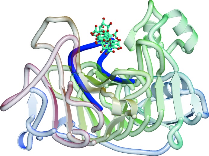
Figure 4.
Model of the binding of the hexasaccharide V of Fries et al. (2007 ▶) in the active site of RW PME. The hexasaccharide is shown in ball-and-stick representation on a ribbon background of the protein. The loop from a superposed YbcH lipoprotein that would interfere with binding of the hexasaccharide is shown as a blue coil.

