Figure 2.
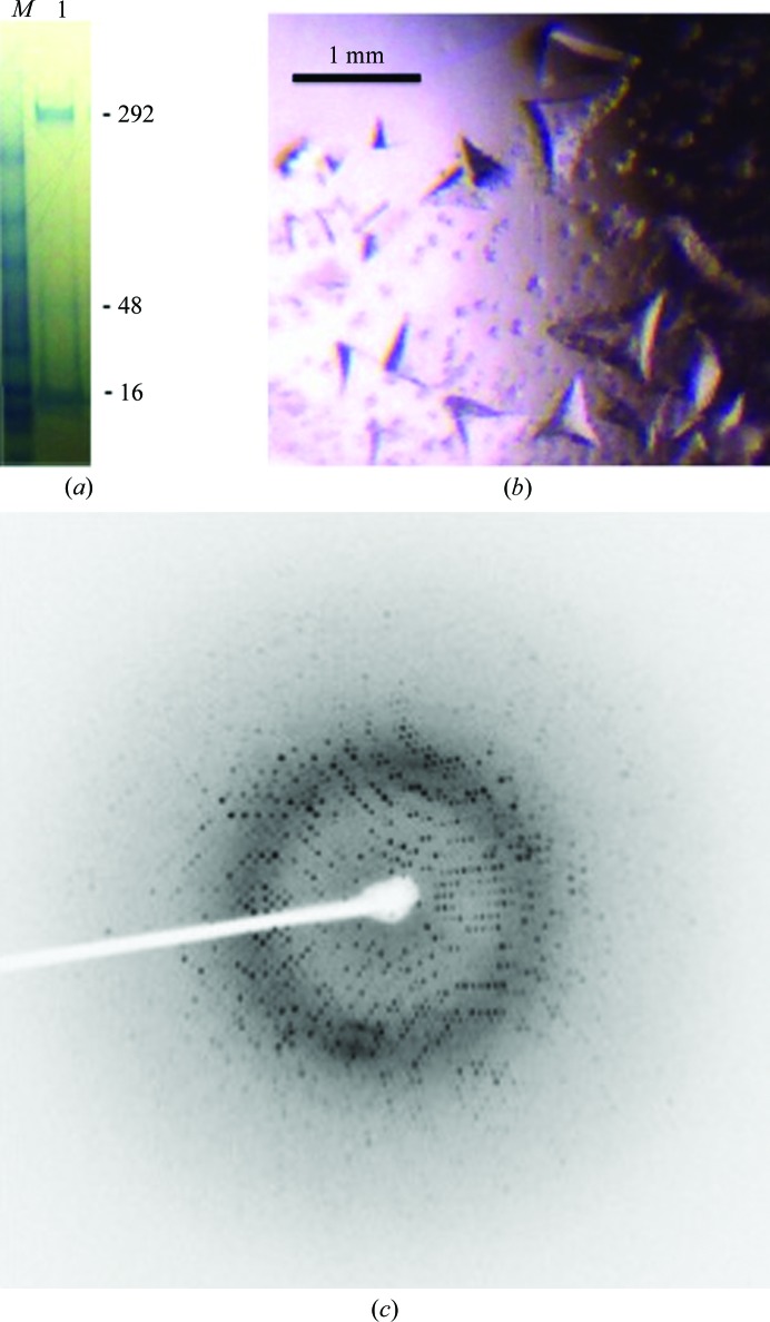
Purification and crystallization of recombinant PaDHQase. (a) Coomassie Blue-stained reduced SDS–PAGE gel of recombinant PaDHQase; the monomeric, trimeric and dodecameric species are indicated by the 16, 48 and 292 kDa molecular-weight markers, respectively. The molecular-weight-marker lane is labeled M, while the protein lane is labeled 1. (b) Crystals of PaDHQase. (c) Sample diffraction image of PaDHQase.

