Figure 3.
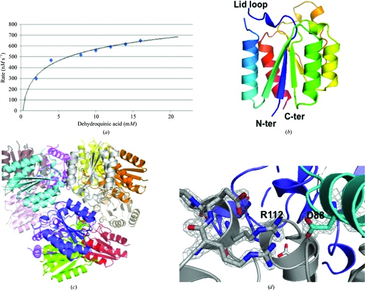
Structure and activity of PaDHQase. (a) Enzymatic activity of 40 nM PaDHQase with increasing concentrations of the substrate 3-dehydroquinic acid. (b) Ribbon diagram of a monomer of PaDHQase rainbow colored from blue (N-terminus) to red (C-terminus); the missing lid loop is also indicated. (c) Dodecamer of PaDHQase with each monomer colored differently. (d) Fit of selected trimer interface residues in the final 2F o − F c electron-density map contoured at 1.6σ.

