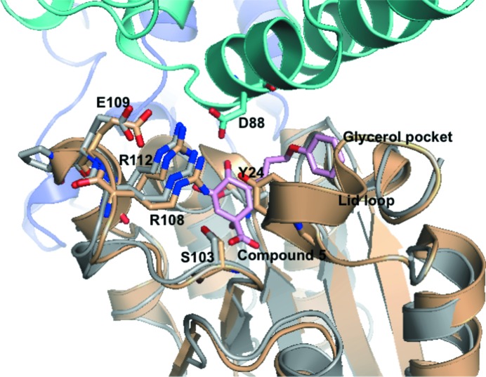
Figure 5.
Comparison of the active sites of PaDHQase and MtDHQase (M. tuberculosis DHQase). The superposed structure of complex of M. tuberculosis DHQase (gold) with compound 5 shows well conserved residues with PaDHQase (shown in stick representation). The location of the lid loop from the MtDHQase structure is also shown with the catalytic tyrosine labeled and shown in stick representation. Key binding-site residues from one monomer of PaDHQase (gray) are in proximity to a helix from another monomer (green). The cavity is large enough to bind compound 5 (in pink stick representation), an inhibitor of M. tuberculosis DHQase that extends into the glycerol cavity. The PDB entry corresponding to the complex of M. tuberculosis DHQase is 3n76 (Dias et al., 2011 ▶).

