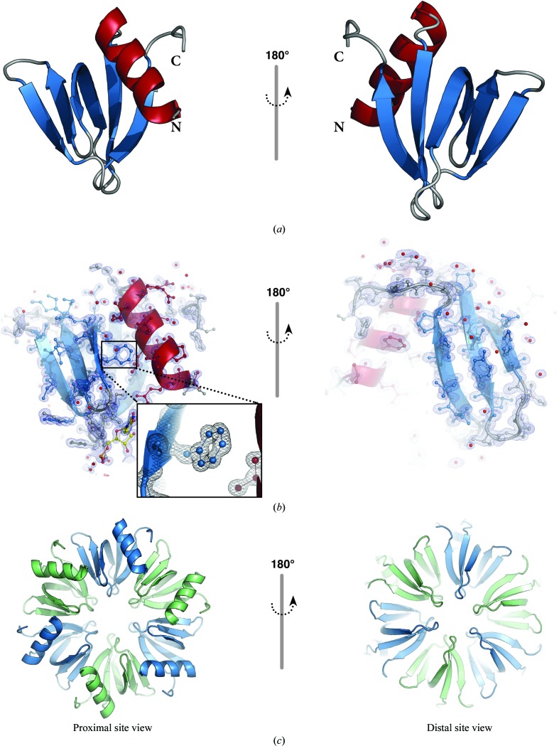
Figure 2.
(a) Cartoon representation of the Hfq monomer present in the asymmetric unit. The single α-helix is coloured red, β-strands are shown in blue and loop regions are shown in grey; the N- and C-termini are indicated. (b) Electron-density map (2F o − F c) contoured at the 1.5σ level. Protein residues and the uridine nucleotide are shown as ball-and-stick representations coloured as in (a) and uridine C atoms are shown in yellow. (c) The sixfold symmetry operation yields the biological assembly, a homohexamer; the individual chains are depicted as cartoons in alternating colours (blue and green).

