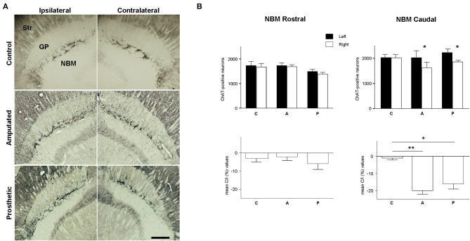
Figure 5.
Cholinergic neurons in magnocellular basal nucleus (MBN). (A) Photomicrographs at low magnification of ChAT-horizontal sections, showing the rostro-caudal extension of MBN in control, amputated, and prosthetic animals. Amputated animals exhibited a significantly lower number of ChAT-positive neurons at the caudal level of the affected side. Prosthetic-animals also showed a significant loss of ChAT-positive neurons in the caudal zone of affected hemisphere. In ipsilateral hemispheres, lateral is left; in contralateral hemispheres, lateral is right; rostral is top. Str, dorsal striatum; GP, globus pallidus; Ret, reticular thalamic nucleus; VP, ventroposterior thalamic nucleus; ic, internal capsule. Scale bar = 500 μm. (B) Estimations of the number of cholinergic neurons in the three experimental groups for the rostral and caudal level separately. Top, inter-hemispheric comparisons (paired t-test, *P < 0.05; **P < 0.01). Bottom, inter-group comparisons (C/I (%), ANOVA *P < 0.05; **P < 0.01).

