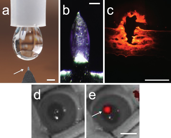
Figure 1.
Microneedles coated with bevacizumab for targeted intrastromal delivery. (a) A single microneedle (arrow) mounted on a handle for manual application is shown next to a liquid drop from a conventional eye dropper. Scale bar: 1 mm. (b) Magnified view of a microneedle coated with bevacizumab. Scale bar: 200 μm. (c) Histological section of rabbit cornea after delivery of AlexaFluor 750–labeled bevacizumab using a microneedle showing the site of microneedle penetration into the eye (arrow) and distribution of red-labeled bevacizumab in the corneal tissue. Scale bar: 1 mm. Image of a live rabbit eye (d) before treatment and (e) after inserting and removing a microneedle coated with AlexaFluor 750–labeled bevacizumab. The red fluorescence indicates the site of bevacizumab deposition within the corneal stroma (arrow). Scale bar: 5 mm.

