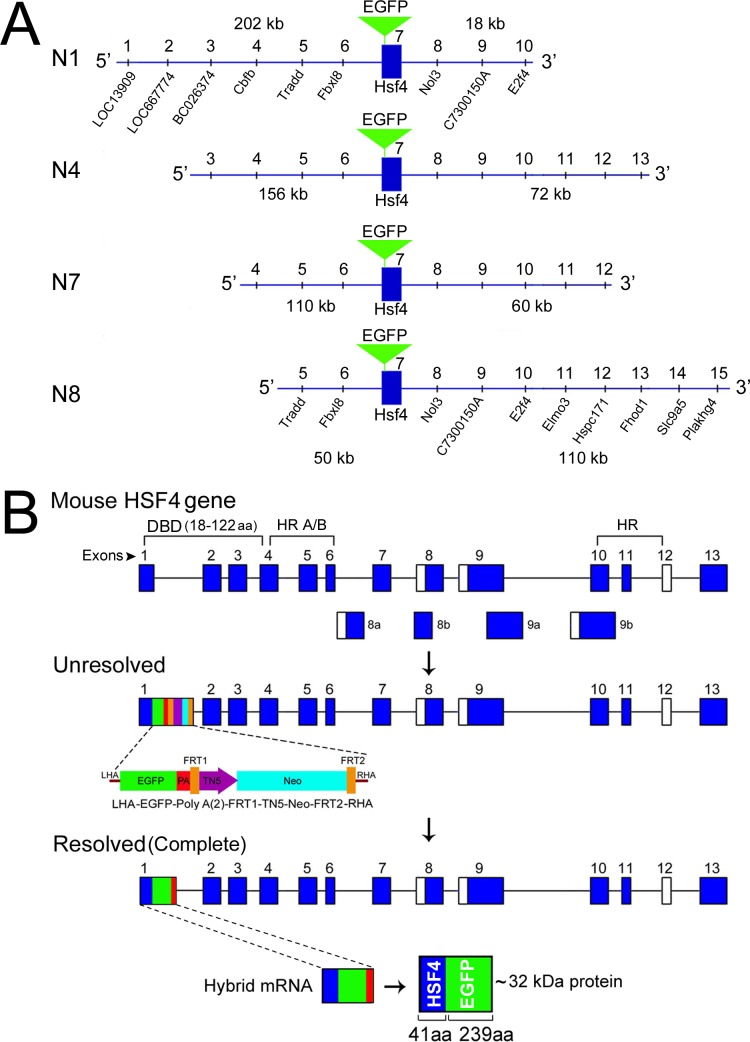
Figure 2.
The Hsf4 manipulated sequences within mouse BACs for expression in the developing ocular lens. (A) N1, N4, N7, and N8 represent four BACs with different lengths of the 5′ (202−50 kb) and 3′ (18−110 kb) surrounding the Hsf4 gene. The common location of the EGFP (green inverted triangle) in the DNA binding domain, at the end of exon 1 of the Hsf4 gene is shown. The markings 1 to 10 designate the positions of the known genes (UCSC Genome Browser). (B) The first schematic shows the Hsf4 gene composed of 13 exons (dark blue rectangles). The first three exons and a part of the fourth exon contain sequences that code for the DNA binding domain (DBD, 104 amino acids long). The location of hydrophobic repeats (HR A/B) is indicated. The open rectangles indicate alternative exons. The second schematic shows the targeting cassette at the end of the exon 1, an intermediate recombineering step in an unresolved BAC. The targeting cassette contains the LHA and RHA (the left and the right homologous arms), the EGFP (green), two polyadenylation signals (PA, red), and neomycin resistance (neo, light blue) driven by TN5 (purple) bracketed by FRT 1 and 2 (brown). The third schematic shows the BAC after recombination is complete (resolved), which removes the TN5-neo-FRT cassette, leaving the EGFP-Poly A sequences attached to the exon 1 of the Hsf4 gene and one FRT site in the resolved BAC (last schematic). The transgene produces the mRNA for the hybrid gene containing the exon1 sequences of the Hsf4 gene (dark blue), the green EGFP coding sequences and the polyadenylation sequence (red). This mRNA translates into an approximately 32 kDa hybrid protein (N-terminal 41 amino acids [aa] of Hsf4-DBD + 239 aa EGFP).

