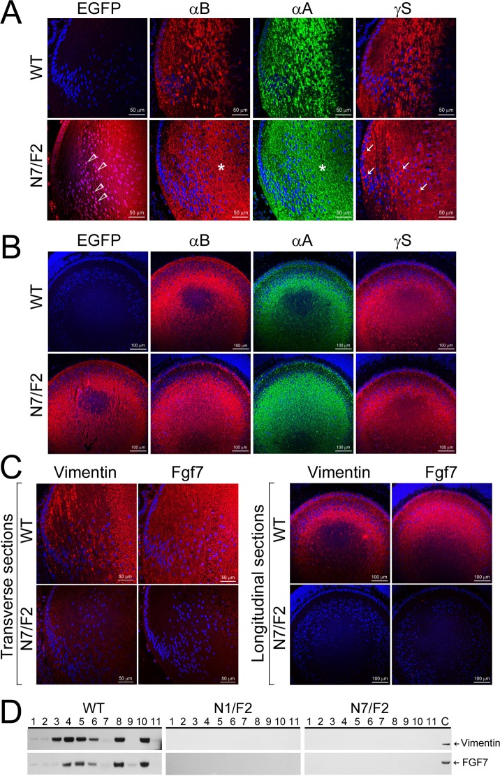
Figure 6.
Altered distribution patterns of αA, αB, and γS, and loss of Vimentin and Fgf 7 expression in PND02 transgenic mouse lens. (A) Immunofluorescence and laser scanning confocal images (transverse/anteroposterior axis) show expression of EGFP (red) in N7/F2 (open arrowheads) and its absence in the WT. The staining patterns of αA (green) and αB (red) show homogenous or granular distribution (asterisks), while γS shows pools of staining (red, possibly aggregated protein, arrows) around the nuclei (blue). Compare this staining pattern with the staining in the first (EGFP) column (N7/F2). The nuclei in the EGFP column are violet (a mix of blue and red, open arrowheads) , we do not see violet nuclei in the γS-stained section, that is because γS is a cytoplasmic protein, while Hsf4(DBD)-EGFP hybrid is a nuclear as well as a cytoplasmic protein. (B) Longitudinal views (perpendicular to anteroposterior axis) of αA, αB, and γS staining in the PND02 lens confirm what is seen in (A). (C) Transverse sections (left) and longitudinal views (perpendicular to anteroposterior axis (right) show loss of vimentin and Fgf7 expression in PND02 transgenic lens. The anteroposterior streaks of vimentin are seen in the WT lens. The staining pattern of Fgf7 is more homogenous all through the cortex in WT lens. (D) Immunoblots showing vimentin and Fgf7 presence in various tissues in the WT mice (1, lens; 2, retina; 3, eye globe; 4, liver; 5, heart; 6, lung; 7, spleen; 8, kidney; 9, small intestine; 10, muscle; and 11, brain; C, control lens extract). However, these proteins are not detected in any of the tissues examined in PND02, N1/F2 and N7/F2 animals.

