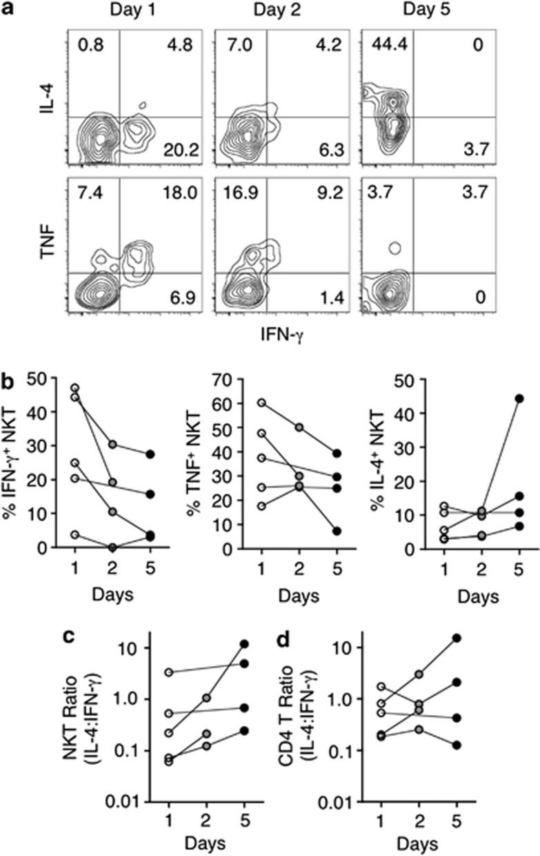
Figure 4.
RIL-21 administration alters the production of T helper 1 (Th1) and Th2 cytokines by natural killer T (NKT) cells. On the days of sample acquisition, an aliquot of cells was frozen for subsequent analysis at a later time. At this time, peripheral blood cells from patients in which NKT cells were most prevalent were stimulated with phorbol 12-myristate 13-acetate and ionomycin for 2.5 h in the presence of GolgiStop. Subsequently, cells were stained for α-GC/CD1d tetramer, CD3, CD4, intracellular interferon-γ (IFN-γ), tumor necrosis factor (TNF), and interleukin-4 (IL-4) and analyzed by flow cytometry. Vehicle-loaded tetramer was also added to the staining cocktail to gate out nonspecific events. (a) Shown are contour plots of NKT cells from one patient over the course of rIL-21 administration. Plots represent IFN-γ vs. IL-4 or TNF on gated NKT cells. (b–d) Graphs depict the total frequency of cytokine-producing cells among NKT cells (b), the ratio of IL-4/IFN-γ production in NKT cells (c) and the ratio of IL-4/IFN-γ production in conventional CD4 T cells (d) from five individual patients.

