Fig. 5.
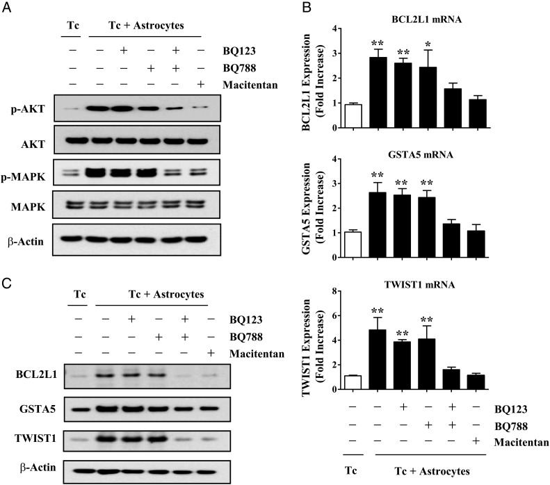
Dual antagonism of ETAR and ETBR block astrocyte-induced activation of AKT/MAPK signaling and anti-apoptotic gene expression in MDA-MB-231 cancer cells. (A) Western blot analysis of MDA-MB-231 cancer cells that were pre-incubated for 2 hours with 1 μM BQ123, 1 µM BQ788, BQ123 plus BQ788, or 100 nM macitentan and then cultured alone (Tc) or co-incubated with murine astrocytes (Tc + astrocytes). GFP-labeled astrocytes were removed by FACS, and Western blot was performed on cancer cells as described in Material and Methods. (B) RT-PCR analysis of BCL2L1, GSTA5, and TWIST1 expression in MDA-MB-231 cancer cells cultured alone or co-incubated with murine astrocytes. Target gene expression levels were normalized to the 18S rRNA level. Fold increase refers to the ratio of mRNA levels in MDA-MB-231 cells cultured alone versus cells co-incubated with GFP-labeled murine astrocytes. (C) Western blot analysis of Bcl2l1, Gsta5, and Twist1expressed by MDA-MB-231 cancer cells alone or cells that were co-incubated with astrocytes for 48 hours. ß-Actin expression was used as an internal control for Western blotting. Values shown are means ± SD of 3 independent experiments, each performed with triplicate cultures. Statistical significances were compared with cancer cell growing alone. *P < .05, **P < .01.

