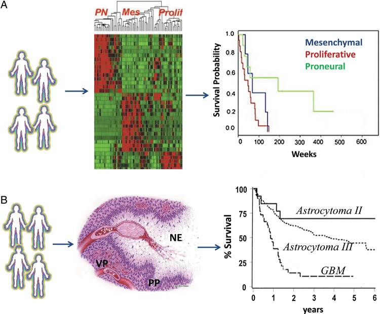
Fig. 1.
Molecular and histolopathological features of GBM. (A) Molecular pathology uses a genomic signature in association with clinical outcomes for diagnosis (adapted from Phillips et al13 with permission). Initial in silico analysis classifies glioblastoma into 3 molecular subsets. A mesenchymal signature indicates a more invasive phenotype. Patients with mesenchymal or proliferative signatures are associated with poor clinical outcome (shorter survival). A proneuronal signature correlates with better prognosis (longer survival). (B) Histopathology relies on morphological changes in association with clinical outcomes for diagnosis. The histological hallmarks of glioblastoma include necrosis (NE), which is often surrounded by pseudopalisading (PP) tumor cells that are migrating away. Vascular proliferation (VP) indicates the rebuilding of the vasculature (adapted from Brat and Van Meir56 with permission). These classic morphological features of glioblastoma correlate with shorter survival (adapted from Prados et al92 with permission).

