Figure 1.
CVP2 Acts Early in Procambial Patterning during Embryogenesis.
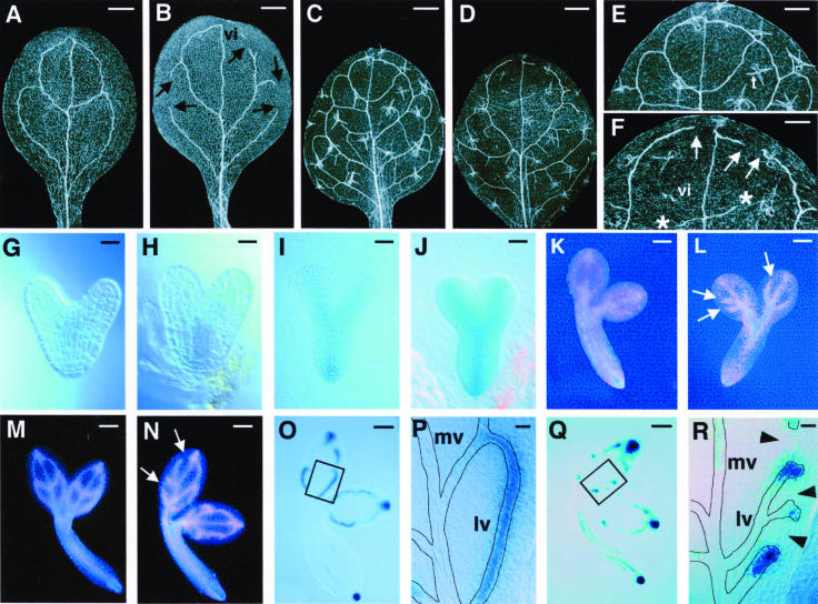
(A) to (F) Cleared specimens viewed with dark-field optics. Wild-type cotyledon (A), cvp2 cotyledon (B), wild-type leaf (C), and cvp2 leaf (D) are shown. Higher magnification of (C) and (D) are shown in (E) and (F), respectively. With dark-field optics, trichomes (t) appear translucent with three spikes.
(G) to (N) Athb8:GUS embryo expression. Wild-type heart (G), cvp2 heart (H), wild-type torpedo (I), cvp2 torpedo (J), wild-type walking stick (K), cvp2 walking stick (L), wild-type bent cotyledon (M), and cvp2 bent cotyledon (N) are shown. Dark-field images are more sensitive than Nomarski optical images and show weak GUS expression as pink and strong GUS expression as blue.
(O) to (R) DR5:GUS expression of bent cotyledon stage embryo. Wild-type embryo (O), wild-type proximal lateral vein at a higher magnification of the boxed region in (O) to show uniform DR5:GUS expression (P), cvp2 embryo (Q), and cvp2 proximal lateral vein at a higher magnification of the boxed region in (Q) to show short stretches of DR5:GUS expression in cvp2 mutants (R). Arrowheads designate absence of DR5:GUS. In wild-type embryos, 369 out of 394 lateral veins showed continuous DR5:GUS expression. In cvp2 embryos, 33 out of 330 lateral veins showed continuous DR5:GUS expression. Roots of embryos in (O) and (Q) were distorted while separating cotyledons. Vein patterns are outlined in (P) and (R).
Arrows indicate free vein endings of secondary veins; asterisks indicate free vein endings of tertiary veins. Dark-field optics were used for (A) to (F) and (K) to (N); Nomarski optics were used for (G) to (J) and (O) to (R). vi, vascular island; mv, midvein; lv, lateral vein. Bars = 500 μM in (A) to (D), 250 μM in (E) and (F), 10 μM in (G), (H), (P), and (R), 25 μM in (I) and (J), 50 μM in (K) and (L), and 80 μM in (M), (N), (O), and (Q).

