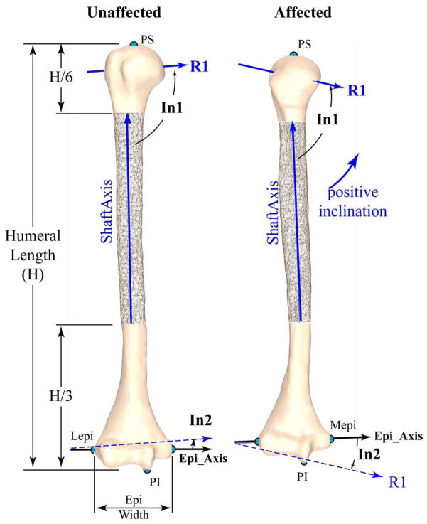
Figure 4. Inclination.
The non-involved left (left) and involved right (right) humerii from a single subject (subject 8, the same as in Figure 3), as viewed in a pure coronal view (after humerii were aligned with the principal axes). A mirror image was created of the non-involved left arm (making it appear as a right arm) for direct visual comparison. Both images are scaled identically. R1, R2, and R3 are the primary, secondary and tertiary radii of the best fit ellipsoid to the humeral head. R1 is shown with an extended length. R2 is in this coronal plane and perpendicular to R1. Both R2 and R3 are not shown for clarity. Inclination is positive when the medial humeral head rotates superiorly. Inclination was the angle between the first principal axis of the best fit ellipsoid to the humeral head (R1) and both the ShaftAxis (In1: head_incl_Shaft) and the epicondylar axis (In2: head_incl_Epi). Lepi and Mepi: most lateral and medial points of elbow epicondylar line; PS and PI: the most superior and inferior points of the humerus. For this subject the measures of inclination, H, and epi_width (non-involved/involved) were as follows: In1: 93.5°/75.7°, In2: 4.1°/−15.0°, H: 241.4 mm/327.4 mm; and epi_width: 63.5 mm/59.2 mm.

