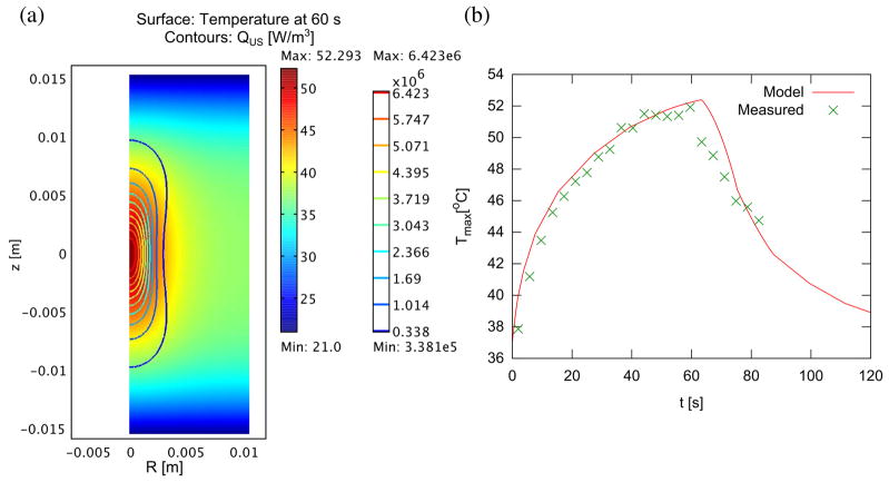
Figure 1.
Temperature distribution in model tissue with cylindrical symmetry (complete domain shown) after 60s sonication (a). Interfaces at z = ±1.5 cm are assumed in contact with room temperature bath, while interfaces at R = 0, 1.5 cm are connected to other tissue, with axial symmetry at R = 0. The ultrasound beam is directed along the R = 0 axis, and is focused at z = 0, with a focal spot size of radius 1.5 mm and length 7 mm. (b) Peak temperature over time during heating and cooling, compared to actual temperature measured during sonication with MR thermometry. Denaturation is assumed to occur between 48 and 50 °C.

