
Fig. 3.
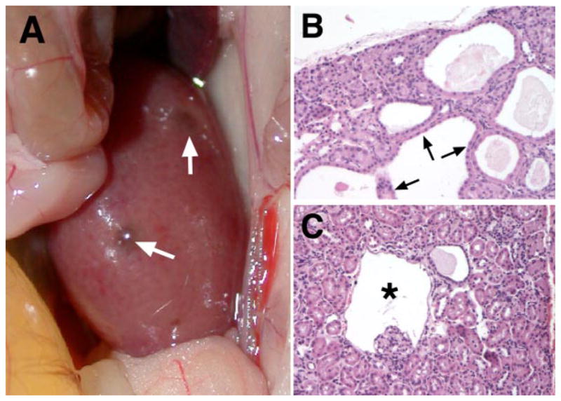
Consequences of von Hippel-Lindau (VHL) gene inactivation in the kidney. Renal cyst development in mice with inactivation of pVHL in proximal renal tubule cells using the PEPCK-Cre transgene (95) is shown. A: macroscopically visible renal cysts in a pVHL-deficient kidney (white arrows). B: multiple renal cysts lined by cuboidal, eosinophilic epithelial cells. Hematoxylin and eosin stain, magnification ×200. C: glomerular cyst development in pVHL-deficient kidneys. Shown is a glomerular cyst (*) with the glomerular tuft located at the cyst basis. Studies with the ROSA26-lacZ Cre-reporter indicated recombination activity in Bowman’s capsule, suggesting that Bowman’s capsule of this cyst is pVHL deficient. Hematoxylin and eosin stain, magnification ×200.