Figure 3.

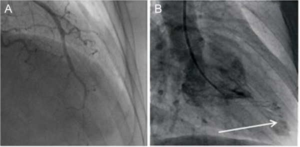
Cardiac catheterization ventriculograms. (A) Image shows chronic occlusion of the distal portion of the anterior descending artery. (B) Ventriculogram of the left ventricle shows an apical pouch or aneurysm (arrow).

Cardiac catheterization ventriculograms. (A) Image shows chronic occlusion of the distal portion of the anterior descending artery. (B) Ventriculogram of the left ventricle shows an apical pouch or aneurysm (arrow).