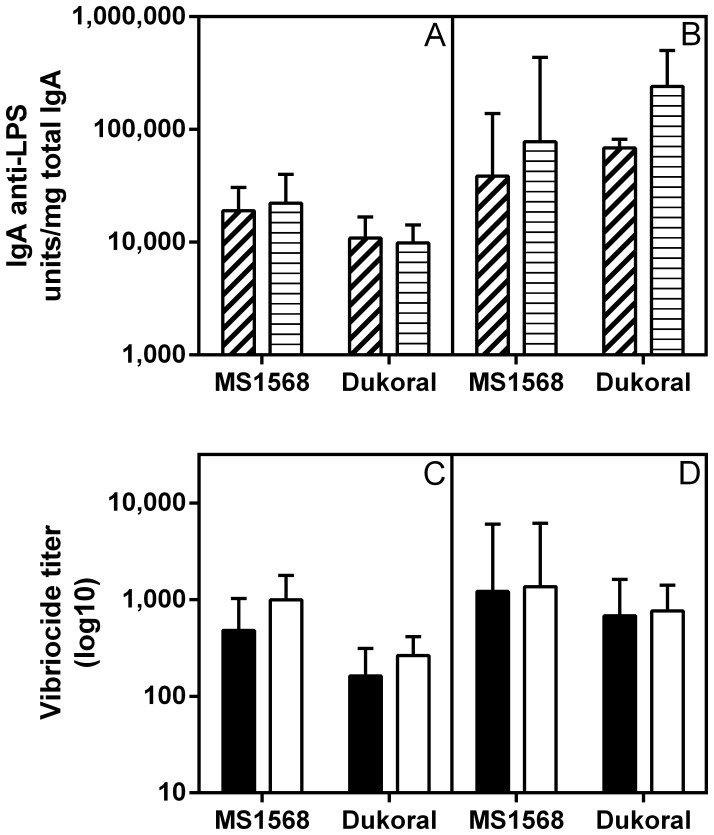
Figure 5. Comparison of intestinal–mucosal IgA anti–LPS and serum vibriocidal antibody responses elicited by oral immunization with formalin–killed MS1568 or Dukoral vaccines in CD1 mice.
(A) IgA anti–LPS antibody levels measured by ELISA in fecal extracts (dashed) and in small intestinal tissue extracts (striped) after two rounds of intragastric immunizations and (B) same after three rounds as described in Material and Methods. (C) Serum vibriocidal antibody responses against Inaba (filled) and Ogawa (open) test organisms after two and (D) three rounds of immunizations. Bars show geometric mean values and SEM for 7 animals per group. As tested with ANOVA, post–immunization antibody levels did not differ significantly between any of the different immunization groups.

