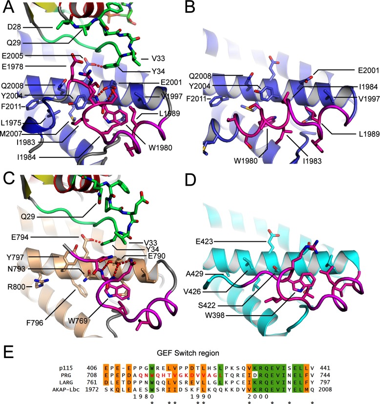
Figure 5. Comparison of the GEF switch region at the N-terminus of the AKAP-Lbc DH domain with the equivalent regions in LARG and p115-RhoGEF.
(A) The conformation of the AKAP-Lbc DH domain GEF switch region (coloured in magenta) when bound to RhoA. RhoA is coloured with red α-helices and yellow β-strands with its switch I region in green. The DH domain of AKAP-Lbc is coloured blue. (B) The conformation of the DH domain when not bound to RhoA. (C and D) The conformation of the equivalent regions in LARG when bound to RhoA (C) and p115-RhoGEF when not bound to RhoA (D). (E) Sequence alignment of the GEF switch regions of AKAP-Lbc and three homologous RhoGEF domains. Residues important for the packing of the GEF switch region are marked with asterisks below the alignment.

