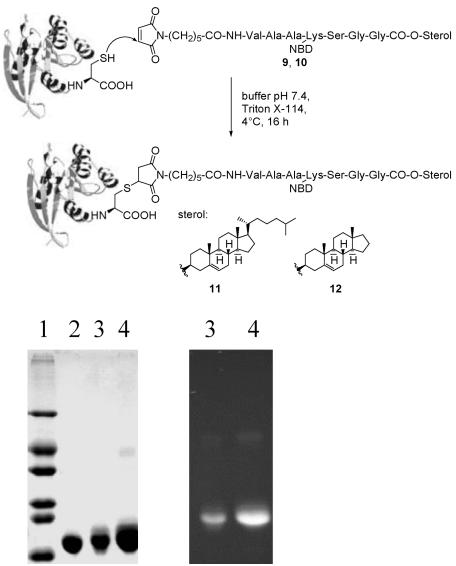
Fig. 3.
Generation of protein chimeras 11 and 12, starting from the lipopeptides 9 and 10. (Upper) Structures. (Lower) Gel electrophoresis of chimeras: after Coomassie-blue staining (Left) and fluorescence (Right). Lane 1, size marker; lane 2, N-RasG12V(1–181); lane 3, N-RasG12V(1–181)-Hh(NBD)-O-cholesterol 11; lane 4, N-RasG12V(1–181)-Hh(NBD)-O-androstenol 12.

