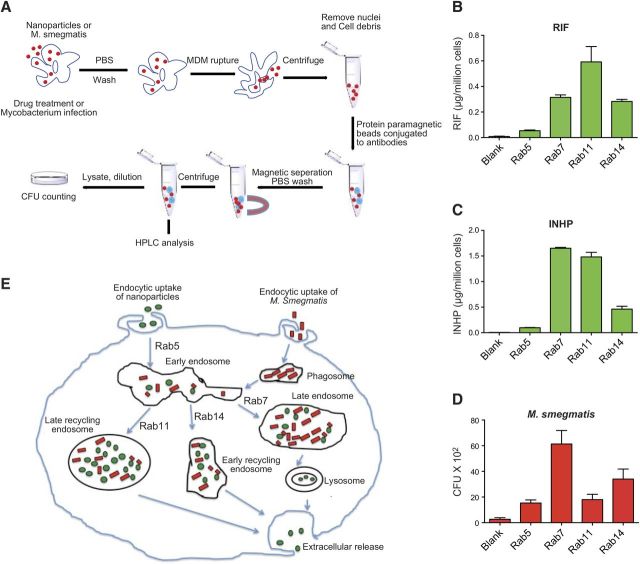
Figure 6.
A) Schematic diagram of immunoisolation of NPs and M. smegmatis-containing endosomes. MDMs were treated with NPs or M. smegmatis side by side. MDMs were then washed in PBS and ruptured in homogenization buffer. Nuclei and unbroken cells were removed by centrifugation. Protein A/G paramagnetic beads conjugated to antibodies were incubated with the supernatants, and the beads containing endosomal compartments were washed and collected on a magnetic separator. Drug content of each compartment was determined by HPLC after sonication. For mycobacterium quantification, the compartments were diluted with sterile PBS (containing 10% human serum), treated with 0.25% SDS, and loaded onto agar plates for CFU counting. B–D) Following immunoisolation, RIF (B), INHP (C), and M. smegmatis (D) were quantitated in subcellular endosomal compartments by HPLC or CFU counting. E) Schematic diagram of intracellular pathways of NPs and M. smegmatis. RIF and INHP NPs (shown in green) and M. smegmatis (shown in red) are phagocytosed by MDMs and transported to early endosomes. Nanoparticles and mycobacteria are then either sorted into late endosomes (Rab 7) for release as secretory lysosome or into fast recycling (Rab14) or slow recycling (Rab 11) endosomes for eventual extracellular release.

