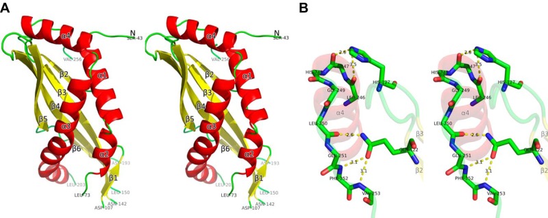
Figure 1.

Structure of SPLUNC1. A) The stereo view of the structure of SPLUNC1 with residues from 43 to 256 (colored with secondary structures). There are 3 missing loops: residues from 74 to 106, residues from 143 to 149, and residues from 194 to 202. All structure figures were prepared by the program PyMOL (http://www.pymol.org). B) Detailed interaction between α4 and 2 conserved residues.
