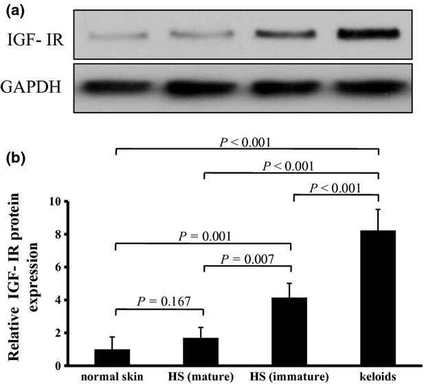
Fig 2.

(a,b) Western blotting analysis of insulin-like growth factor-I receptor (IGF-IR) protein expression in cultured fibroblasts of keloid, immature hypertrophic scar (HS), mature HS and normal skin. (a) Cell lysates from fibroblasts were prepared and subjected to Western blotting with antibodies against IGF-IR and glyceraldehyde 3-phosphate dehydrogenase (GAPDH). (b) Mean densitometric data showing the level of IGF-IR protein normalized to that of GAPDH protein. Data are expressed as mean ± SD (n = 3). Statistical analysis was performed by ANOVA followed by Bonferroni-corrected independent t-tests. ANOVA = 0.000. P < 0.0083 was considered statistically significant.
