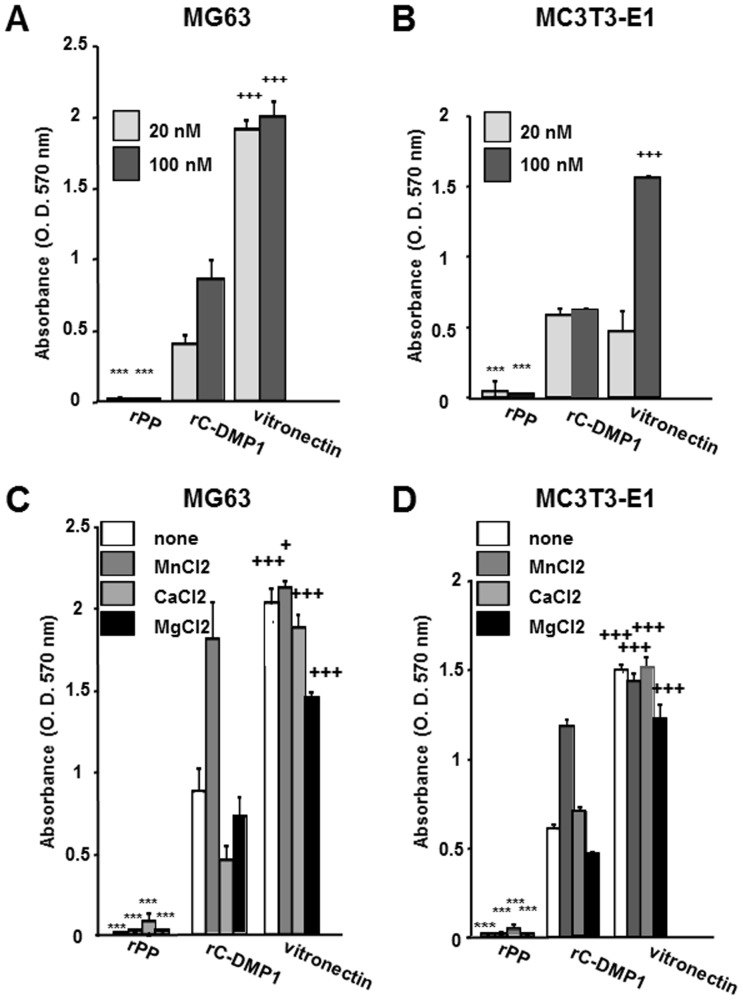
Figure 3. Nominal effects of rPP on MG63 and MC3T3-E1 cell adhesion.
Ninety-six-well plates were precoated with 20 or 100 nM rPP, rC-DMP-1, and vitronectin, seeded with MG63 (A) and MC3T3-E1 (B) cells in serum-free medium, and incubated for 1 hr. MG63 (C) and MC3T3-E1 (D) cells were seeded onto 100 nM rPP, rC-DMP-1, and vitronectin with either MnCl2, CaCl2, or MgCl2 (1 mM) in serum-free medium, and incubated for 1 hr. After washing non-adherent cells, the attached cells were stained with 0.2% crystal violet and dissolved in 1% SDS solution. Absorbance was measured at 570 nm. Each value represents the mean of triplicate determinations; bars mean ±SD. Statistical analysis was performed by a one-way ANOVA, followed by Dunnett's test. ***p<0.001 indicates significantly lower and +p<0.05 and +++p<0.001 indicate significantly higher than rC-DMP-1-coated wells at the same concentration (A and B) or rC-DMP-1-coated wells with the same divalent cation (C and D).

