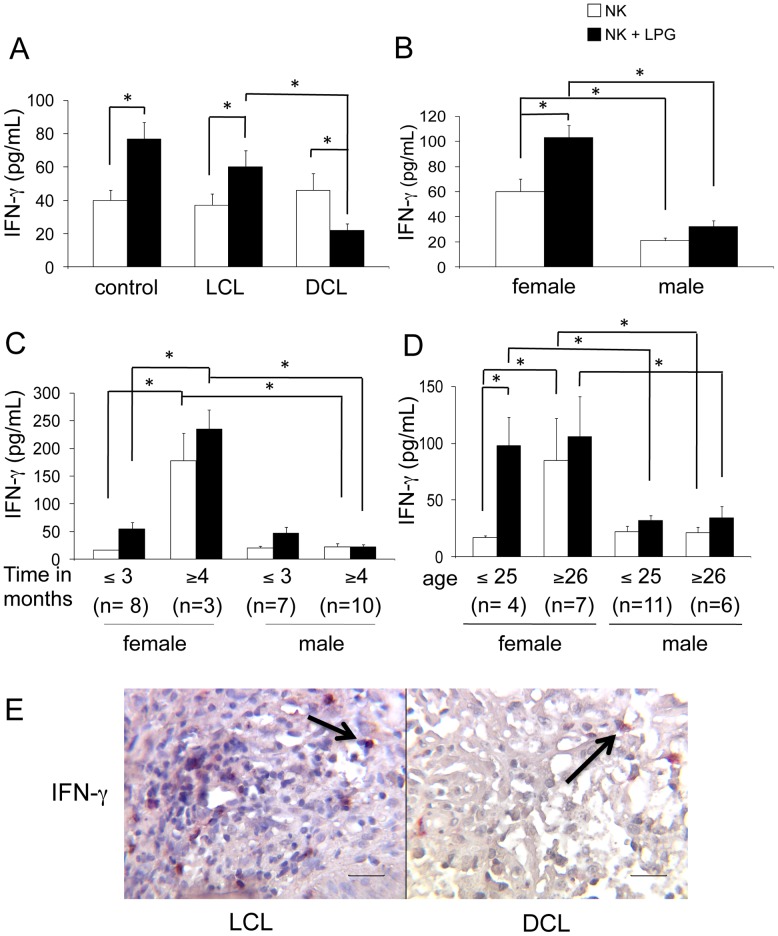
Figure 8. IFN-γ production by NK cells.
(A) IFN-γ production of peripheral blood NK cells [control subjects (n = 21), LCL patients (n = 28) and DCL patients (n = 6)]. Analysis in LCL patients [female (n = 11) and male (n = 17)] according to: (B) gender; (C) disease duration; (D) age. (E) Double immunohistochemical labelling (CD57+/IFN-γ+) in lesions of patients (LCL and DCL) showed redish-brown staining generated by the combination of a red AP substrate used for NK cells and DAB Black used for IFN-γ staining. □ Non-stimulated NK cells. ▪ LPG-stimulated NK cells. Mean ±SEM is shown. *p≤0.05 was considered significant. Scale bar = 50 µm. Black arrows show double positive cells.

