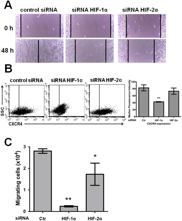
Figure 4. Stable knockdown of both HIF-1α and HIF-2α decreased the cell migration of SW480 cancer cells, but only HIF-1α knockdown severely affected CXCR4-mediated chemotaxis.
A) A wound-healing assay was used to determine whether cellular migration is dependent on either HIF-1α or HIF-2α expression. SW480 knockdown and control (scrambled shRNA plasmid) cells were grown to confluence on 24-well tissue culture plates, and the wound-healing assay was performed as described in the “Material and Methods”. The scratched area was imaged immediately after wounding (time 0) and 48 h after wounding. These images are representative of three independent experiments. B) CXCR4 expression decreased as a result of HIF-1α knockdown. SW480 control or silenced cells, as indicated in the figure, were detached using EDTA and washed, and 1×10⁵ cells were incubated with mouse biotin-conjugated anti-CXCR4 and allophycocyanin-conjugated streptavidin antibody and examined by flow cytometry. The data show the median fluorescence intensities of CXCR4 expression and SSC (side-scattered light, proportional to cell granularity), and represent the mean values ± SEM from three independent experiments. *: p<0.05; **: p<0.01. C) Significant differences were observed in the SDF-1α-induced chemotactic response. The chemotactic response to SDF-1α of the HIF-1α- or HIF-2α-knockdown or control cells (Ctr in the figure) was assessed using Boyden chambers as described in the “Materials and Methods”. The cells were incubated for 48 h to allow for migration and recovered using EDTA. The results represent the means ± SEM from three experiments performed in duplicate. *: p<0.05; **: p<0.01.

