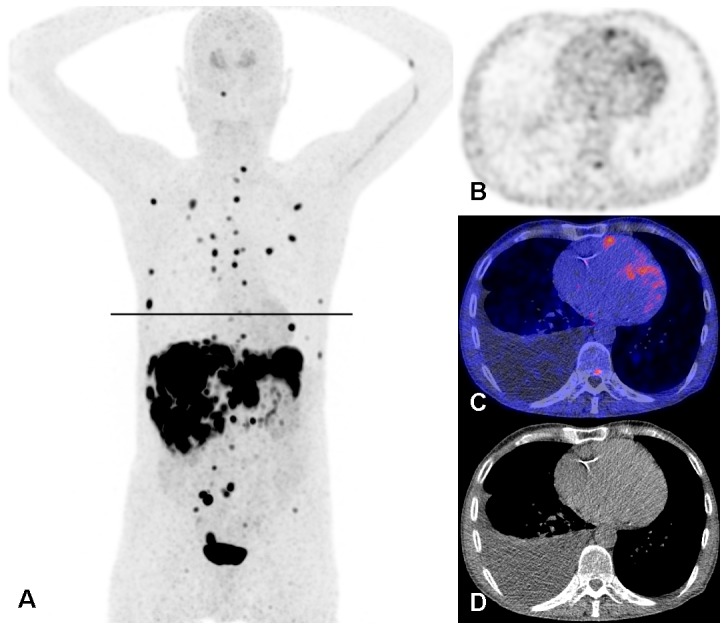
Figure 4. 18F-FDOPA PET/CT scan of the patient illustrated in Fig 2 .

A: Maximum intensity projection showing multiple metastases of the carcinoid tumour in the mediastinum, thoracic wall, liver, mesentery and skeleton. Physiological uptake in the striatum. Administration artefact in the left elbow and vein in the left upper arm. The black line is the slice position of panels B, C, and D. PET alone (B), fused PET/CT (C) and CT alone (D) images on the slice position indicated in panel A. Physiological uptake in the myocardium with a focus of increased uptake in the right ventricle wall. Also small focal uptake in the posterior part of a thoracic vertebral body, right sided pleural effusion and atherosclerosis of the right coronary artery.
