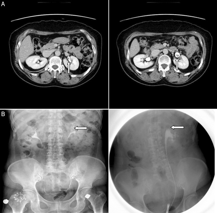
Figure 2.
(a) A fibroepithelial polyp in the left renal pelvis in a 55-year-old woman who visited our clinic for further evaluation of a 1.6-cm left renal pelvis mass that was detected incidentally during an attempted workup for gallbladder stones at a local clinic. An excretory-phase scan shows a 1.6-cm ill-defined mass (arrow) as a filling defect in the left renal pelvis. (b) Both the intravenous pyelogram (left) and the retrograde pyelogram (right) show the focal filling defect (arrow) in the upper calyx portion of the left renal pelvis.

