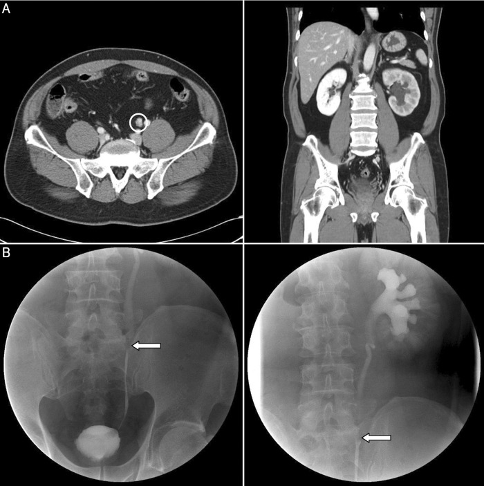
Figure 3.
(a) An inflammatory pseudotumor in a 49-year-old man with vague left flank pain. A parenchymal phase scan shows a 1.5-cm well-enhancing mass in the left midureter with nearly total obstruction causing hydroureteronephrosis (left, circle). In the coronal image, the left kidney has moderately decreased perfusion of the renal parenchyma, which suggests that its renal function is slightly reduced compared with that of the right kidney (right). (b) A retrograde pyelogram shows dilation of the ureter and renal pelvis distal to the obstructing tumor (arrow), namely, the “goblet sign.”

