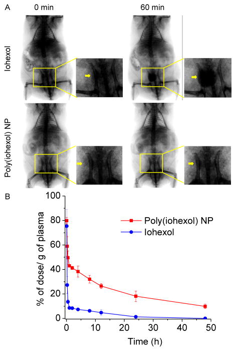
Figure 3.
(A) Serial fluoroscopic images of C57BL/6 mice following jugular vein injection of 200 μL of conventional iodinated contrast agent (iohexol) solution (upper panel) and poly(iohexol) NP solution (lower panel) at 250 mg iohexol/kg, respectively. Images taken at 0 min and 60 min after injection were shown. Arrows indicated the enhanced contrast in the bladder regions. (B) In vivo circulation time of poly(iohexol) NP and iohexol. 64Cu labeled poly(iohexol) NP and iohexol were injected intravenously through the tail vein of mice. At various time points (5 min, 15 min, 30 min, 1 h, 2 h, 4 h, 6 h, 8 h, 12 h, 24 h, and 48 h), blood was withdrawn intraorbitally and the radioactivity was measured by the γ-counter to evaluate the systemic circulation of the poly(iohexol) NP (red) and iohexol (blue) (n=3).

