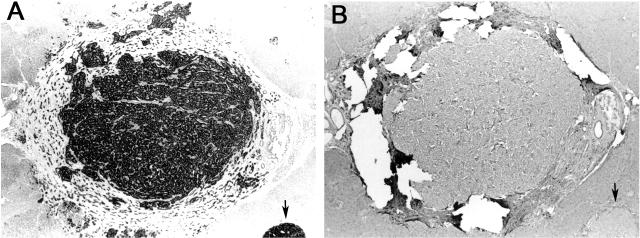
Figure 3. Lack of FLM staining of an MEN-1 glucagonoma with germline mutation K119del known tumor LOH.
Panel A - Centrally located tumor surrounded by a thick fibrous capsule and a smaller tumor at the low right corner (arrow) are strongly positive for glucagon (original magnification ×40). Panel B - Consecutive histological section stained with FLM demonstrates lack of immunoreactivity in both tumors. Note - tears in the section resulted from rupture of the thick fibrous tumor capsule during the antigen retrieval procedure (FLM at 1:300, original magnification ×40).

