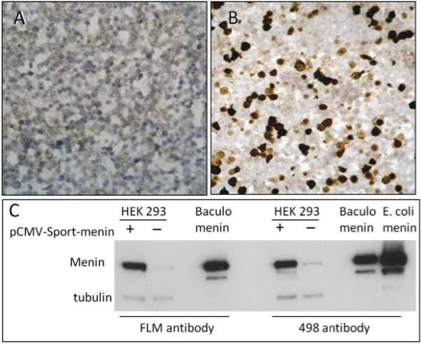
Figure 4. FLM staining results in HEK 293 cells.

Panels A, B – Histological sections from an HEK 293 FFPE cellblock stained with FLM antibody show no immunoreactivity in untransfected cells (A) and strong nuclear staining in cells transfected with full length MEN1 mRNA (B) (Original magnification x400). Corresponding immunoblot results with the FLM and 498 antibodies and controls are shown in panel C.
