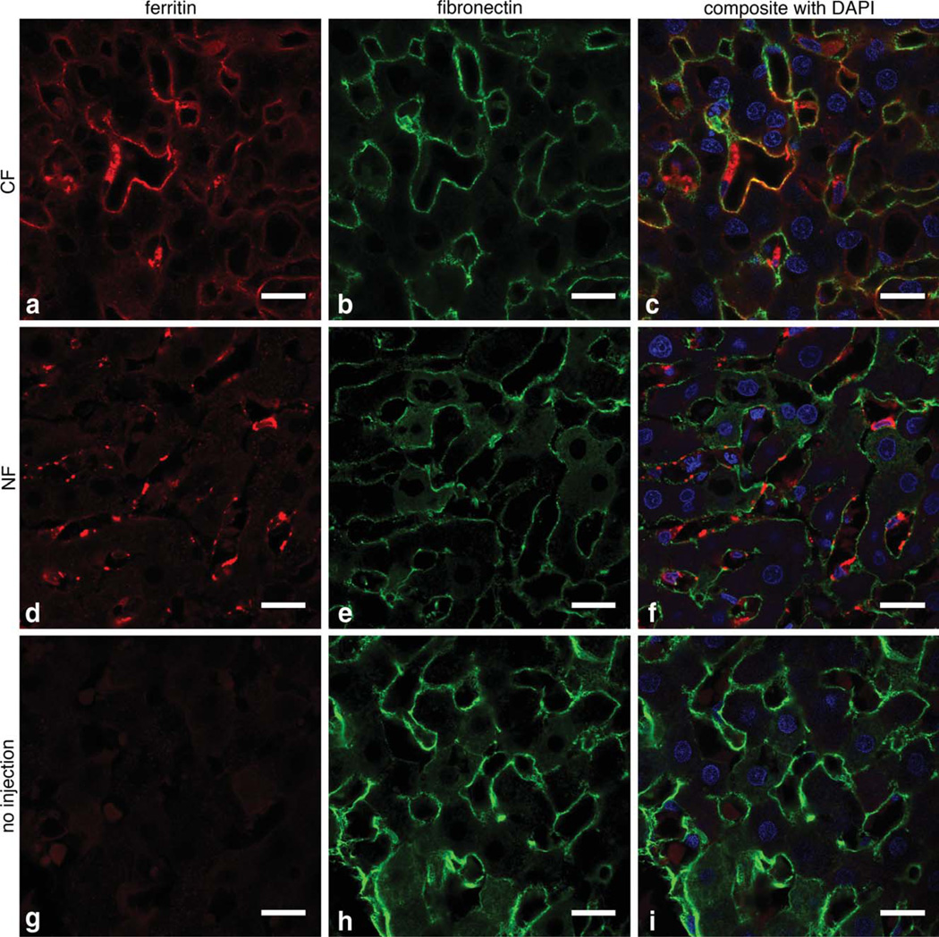
FIG. 2.
CF labels the perisinusoidal ECM 1.5 h after intravenous injection, as shown in 63 × IF imaging. a–c: Show that CF (red) and fi-bronectin (green) are co-localized, consistent with ECM labeling with CF. No co-localization of NF with fibronectin is observed (d–f); instead NF appears to be taken up into macrophages and endothelial cells. Minimal ferritin IF was observed in the unlabeled control liver (g–i). Scale bar = 20 µm, cell nuclei are stained with 4’,6-diamidino-2-phenylindole (blue).

