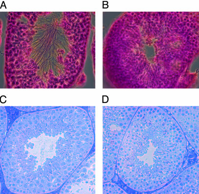
Fig. 4.
Histological examination of mouse seminiferous tubules. (A) Hematoxylin and eosin staining of a testes section from a Bbs4+/+ mouse. Flagella are apparent in the lumen of the tubule. (B) Hematoxylin and eosin staining of a testes section from a Bbs4–/– mouse showing the absence of flagella in the lumen. (C) Toluidine blue staining of a 1-μm testes section from a Bbs4+/+ mouse showing spermatozoa with normal morphology as evidenced by the presence of flagella and condensed sperm nuclei. (D) Toluidine blue staining of a 1-μm testes section from a Bbs4–/– mouse showing the presence of condensed sperm nuclei in the absence flagella.

